Field work got messy last week (this is a warning for messy pictures below).
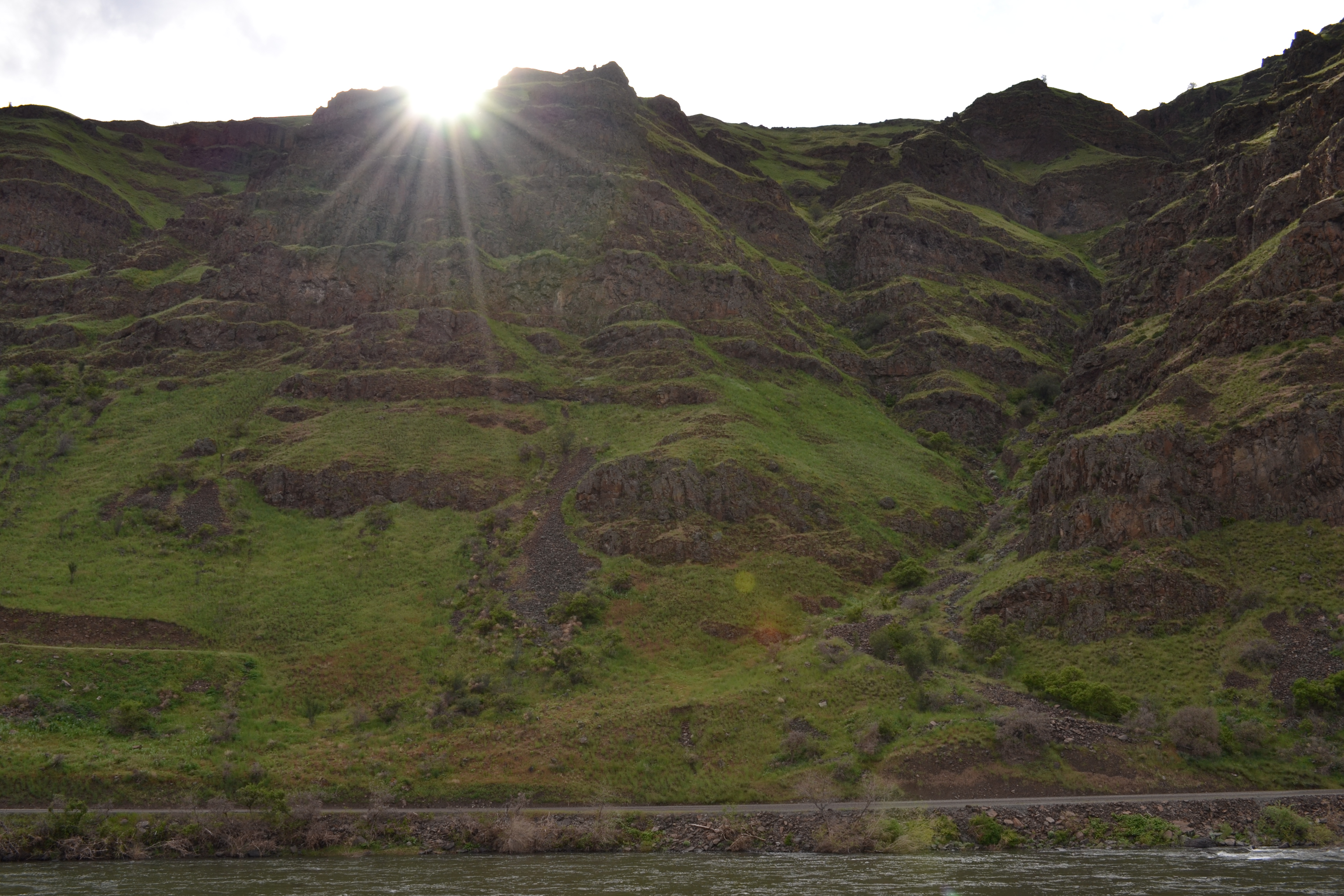
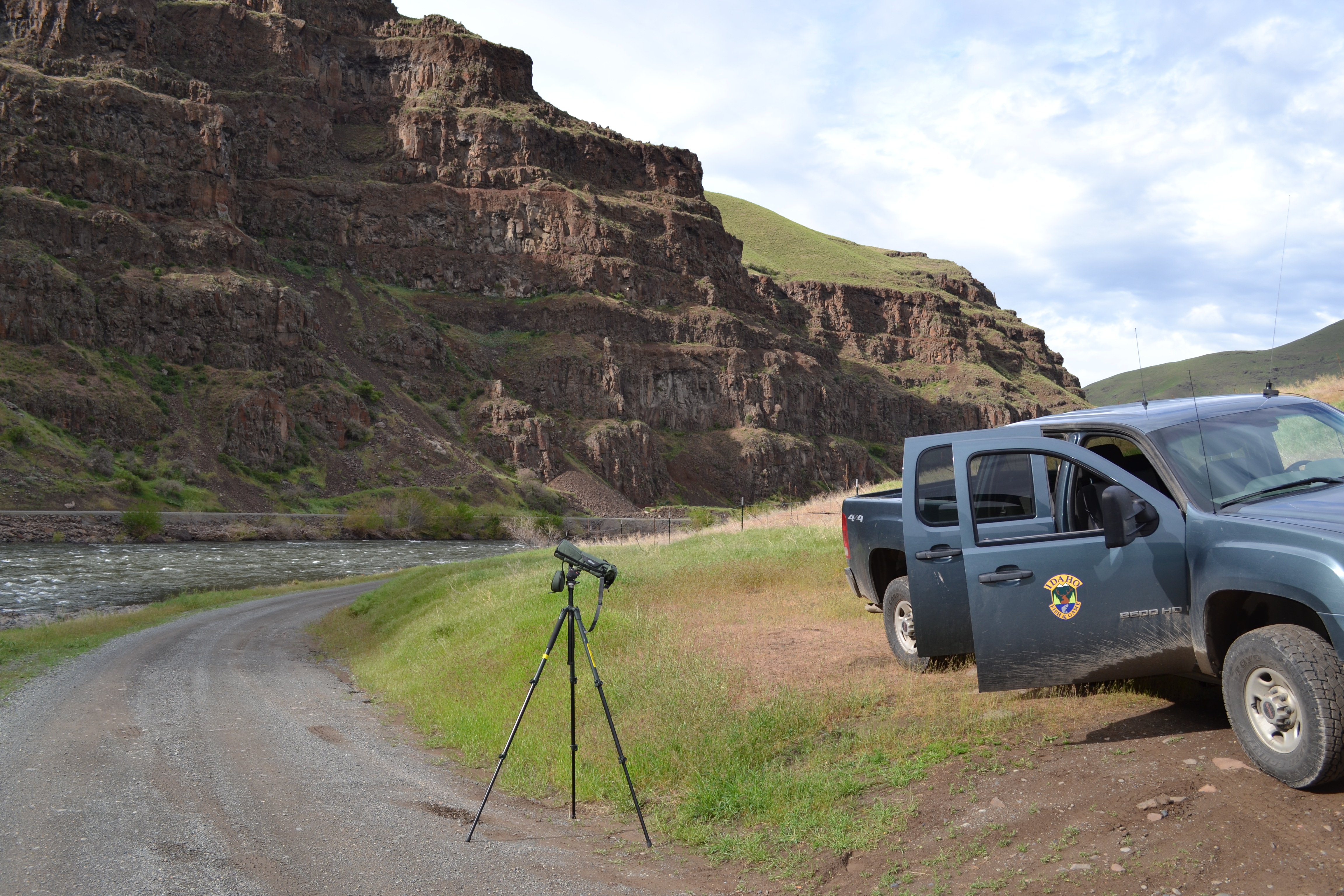
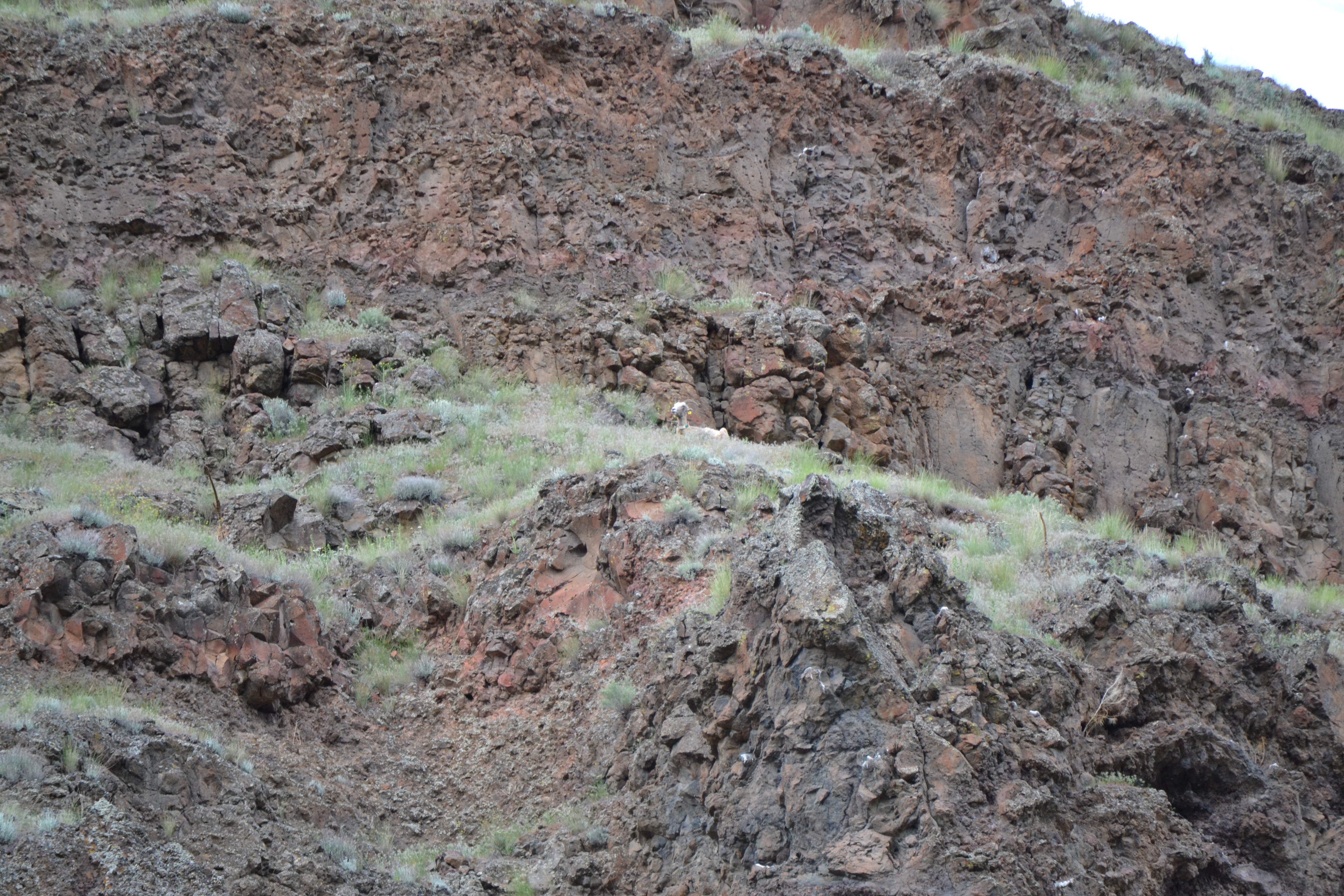
Most days of working in the field start out the same: Kezia and I wake up, drive through canyons with our radio receiver and telemetry equipment to listen for sheep and scan hillsides for visual cues of ewe groups. But on Sunday, we strayed from the normal routine of collecting observational data when our scopes hit something we’ve been both expecting and fearing: our first dead sheep.
Working amidst an outbreak, death is expected. Disease kills. We recognize this when we talk about disease imposing selection pressure, when we talk about host-pathogen evolution, or when we talk about virulence-transmission trade-offs. Death is different when it looks like a dead sheep on a hill side.
What do you do with a dead sheep? Kezia and I were told that we needed to recover the carcass and bring it to the vet lab where necropsy experts could determine the cause of death and microbiologists could test for the presumed causative agents of pneumonia. A carcass contains valuable data in an epidemic setting, especially in a system with a small population size, difficult terrain to navigate and rarely observed fresh carcasses (in remote areas, carcasses are sometimes not found for 5 to 10 days, if not months postmortem when body conditions are close to clean bones). We were told to find the carcass, hike it back to our truck and then deliver the whole body to the vet school in Pullman.
What do you do with a dead sheep when you can’t carry it back to your field truck? By the time we found additional hands to help carry the sheep, and managed to hike our way to where the ewe died, she had been dead for 40+ hours. Her carcass, sitting on a cliff in the middle of a hot Hells Canyon, was colonized by maggots. The skin sloughed off when we lifted the body. One-hundred-fifty pounds of dead weight needed to come down an incline that took both hands to boulder up. We could not carry the whole body out of the canyon, at least not in one piece.
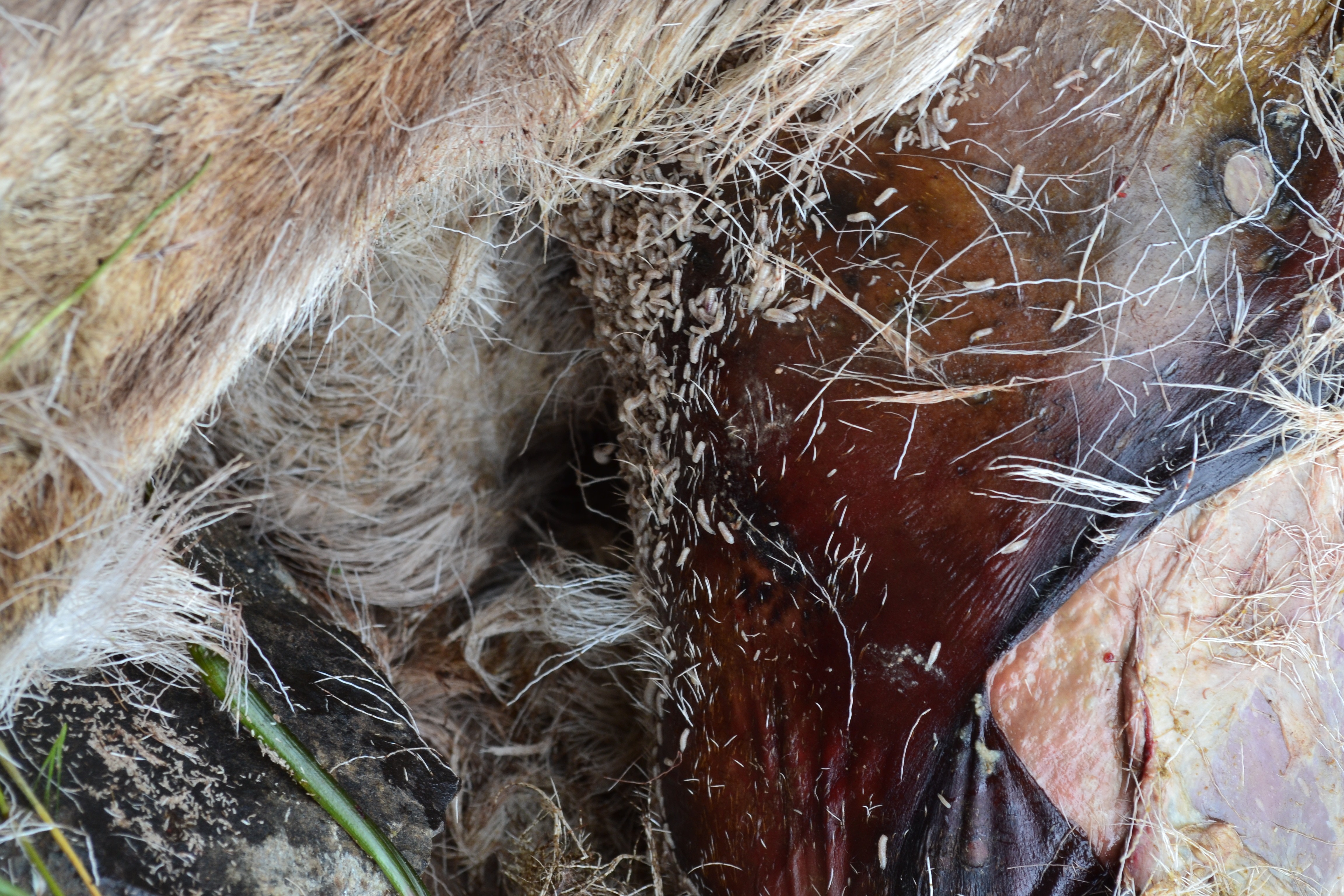
Maggots break down tissues quickly postmortem. Forensic entomologists can use images like this one to gauge how long a carcass has been in an area and estimate time-since-death.
Option B was to do a necropsy in the field and bring back the essential parts which were more feasible to carry: the head and the pluck. For pneumonia, the most important samples come from the lungs, trachea and respiratory tissues.
I had never done a field necropsy. I had never touched a sheep. I was supposed to bring back the pluck. What was “the pluck”*?
Lucky for Kezia and me, one of the field biologists who tracks the bighorn population across the river from where we work had done a necropsy before and was willing to show us how it is done.
This is how you do a field necropsy:
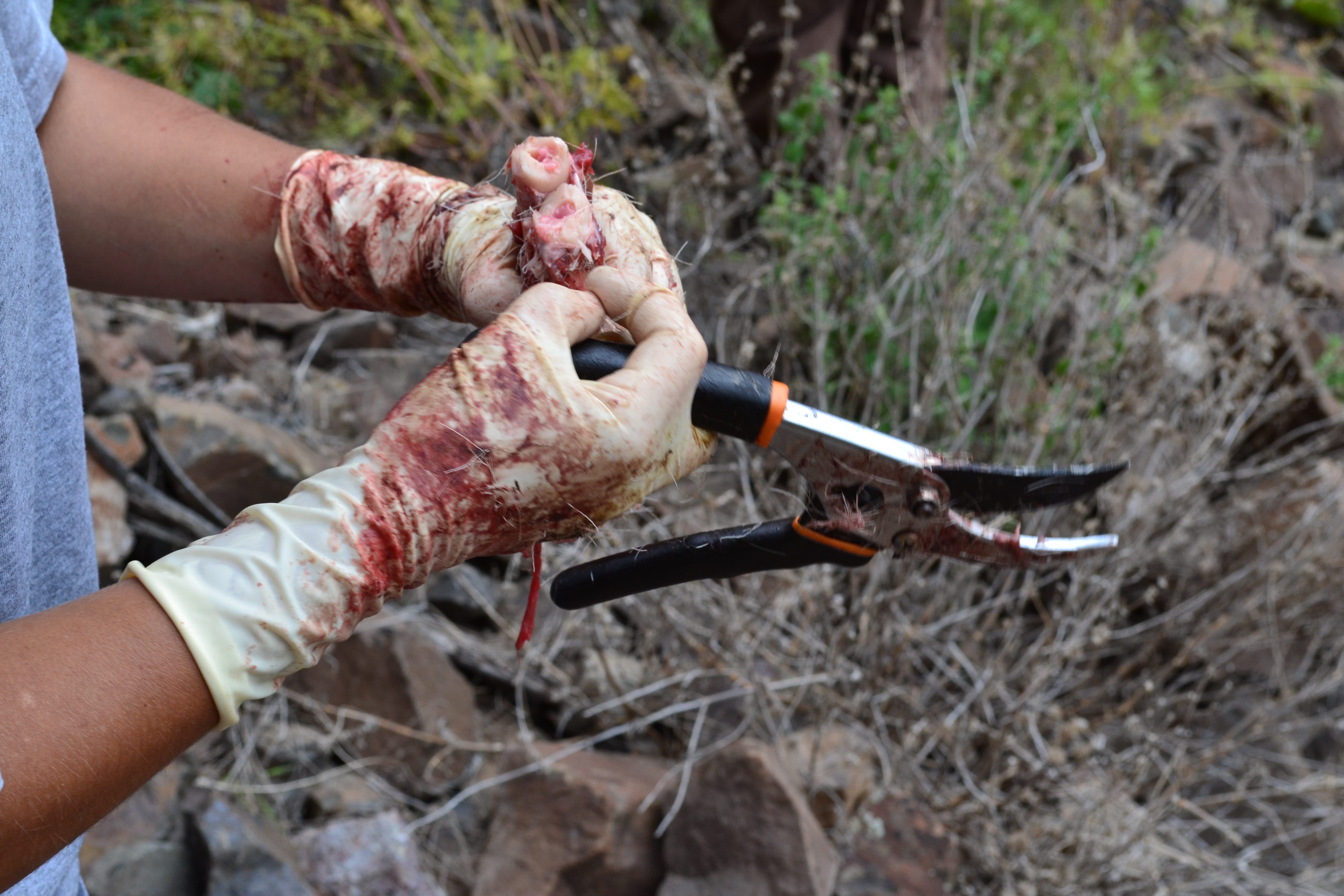
Step one is to remove the head and try to take the pluck out intact. You do this with tools included in a “necropsy kit” — the kit includes a large knife, a saw, pliers, sampling bags, gloves, and a cutting scissor-like tool that reminded me of pruning sheers (you use those to crack ribs if the lungs don’t slide out).
Step two is to take out other organs which may not have come out with a first attempt to slide out the pluck. Bodies decay quickly in summer sun so we needed to remove additional parts of the lungs which were not removed with the trachea and upper respiratory tract (the lungs had nearly liquefied).
Step three is to take proxy measurements of the sheep’s health status. You do this by looking at the bottom of the hooves and checking the color of the bone marrow. With the hooves, you are checking to see if there are any abnormal growths, a common indicator of poor health in bighorns. The color of the bone marrow is used to gauge nutritional status. White bone marrow is healthy and rich in fats, indicating that the sheep had high enough body fat that it would not have died from starvation. Red marrow indicates that a sheep has used up all of it’s bodily fat reserves and has begun to use it’s last reserves — the fat reserves in the bone marrow. Our sheep had nice white marrow.
The inside of a femur bone gives us a glimpse of marrow, which here looks healthy as indicated by the white to pinkish color.
Step four is to bring back the samples to a lab where other scientists can test for pneumonia pathogens and give us a better idea of the cause of death. Even if the ewe did not die of pneumonia, testing the respiratory tissues for pneumonia agents will give us an idea of whether she had been infected in the past or was harboring low densities of pathogens that may not have made her sick but could have allowed her to spread the infection to sheep in her ewe group or to young lambs in the same group.
The data we obtain from carcasses will give us a better idea of the prevalence of infection in the population and the mortality rate associated with pneumonia. When we recover samples like we did on Sunday we hope that we are working toward a better understanding of how, when and why the outbreaks occur. The experience has left me so far with a deep appreciation of data from wildlife systems: often we look at a data set and see points on a graph. Out here, I am reminded of where mortality statistics come from and what disease as an evolutionary selection pressure looks like “on the ground.”
*the pluck is a term used to refer to internal organs